Tube Shunt Surgery
A tube shunt, also called a glaucoma drainage device or implant, is a small silicone tube connected to a thin plate. The tube is placed just inside the front of the eye and carries fluid back to the plate, which sits on the white of the eye under the eyelid, well behind where it can be seen. Fluid collects in a reservoir around the plate and is absorbed. Common devices are the Ahmed valve and the Baerveldt and ClearPath implants.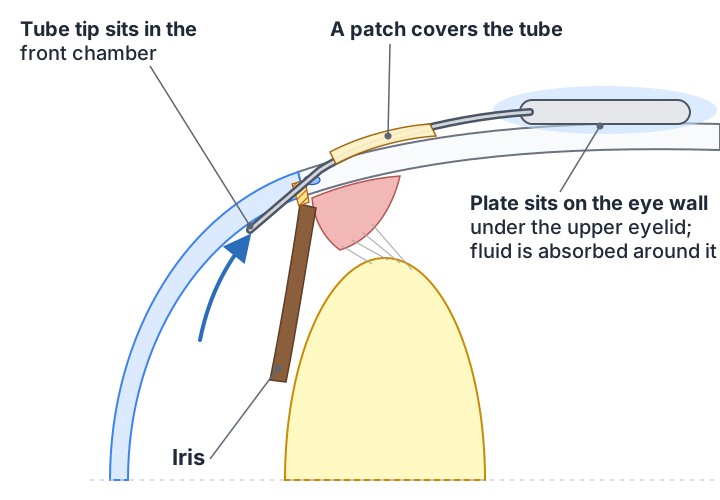
Who it is for
Tube shunts lower pressure about as well as trabeculectomy and are chosen when a trabeculectomy is less likely to work or has already failed. That includes eyes with scarring from previous surgery or injury, glaucoma caused by inflammation or abnormal blood vessels (neovascular glaucoma), some eyes that have had corneal transplants or retinal surgery, and eyes where very close early follow-up would be difficult. In many practices, tube shunts are now used as a first surgery for selected patients.
Valved and non-valved devices
An Ahmed implant has a valve that opens at a set pressure, so it begins working right away and the risk of pressure dropping too low is less. Baerveldt and similar implants have no valve; the surgeon ties off the tube with a dissolving stitch so it does not drain for the first four to six weeks while a capsule forms around the plate. During those weeks pressure is controlled with drops, and when the stitch dissolves the tube opens. Non-valved devices tend to reach slightly lower pressures in the long run. Your surgeon will explain which is planned for you and why.
The day of surgery
Surgery is at an outpatient center under a local anesthetic block with sedation, and takes about an hour. The plate is secured to the eye wall under the conjunctiva, the tube is passed into the front of the eye through a tiny tunnel, and the exposed part of the tube is covered with a patch of donor tissue or your own sclera so it does not wear through. You go home the same day with a patch and shield and need a driver.
Recovery
Drops. A steroid drop is used frequently at first and tapered over about six weeks, an antibiotic for the first week, and an antibiotic ointment at bedtime for about two weeks. Use the Post-Op Eye Drop Schedule tool to print the day-by-day plan. With a non-valved implant you will also keep using some glaucoma drops until the tube opens; your surgeon will tell you which.
Visits. Next day, one week, then every one to two weeks for the first six weeks, then at increasing intervals. With a non-valved device there is often a visit around the time the tube is expected to open.
What is normal. Blurry vision, a scratchy feeling, mild ache and a red, swollen eye for the first one to two weeks. Vision gradually improves over one to two months. The eye may feel slightly "full" on the side where the plate sits. Occasionally there is a feeling of double vision early on from swelling around the muscles; this usually settles.
Activity. Shield at night for two weeks. For the first month, avoid rubbing the eye, swimming, heavy lifting and bending with the head down. Reading and screens are fine.
Call the same day if
- Pain increases rather than eases
- Vision suddenly worsens
- The eye becomes much redder or has discharge
- You can see or feel the tube, or the eye feels very hard or very soft
Results and risks
Most eyes reach target pressure, with or without drops, and the implant lasts for life. Pressure may drift up over the years as the capsule around the plate thickens; this is usually managed with drops.
Risks include pressure that is too low or too high in the early weeks, bleeding or inflammation inside the eye, double vision, the tube touching the cornea or being too long or short (correctable), and the covering over the tube wearing thin over years, which can be repaired. Gradual clouding of the cornea is a recognised long-term concern that your surgeon monitors. Infection is rare. As with any glaucoma surgery, lost vision from glaucoma is not restored; the aim is to protect what remains.
Questions worth asking
- Valved or non-valved, and why?
- Which of my drops do I continue after surgery?
- When should pressure be at target?
- What would make you want to see me urgently?