Retinal Vein Occlusion
Published
The retina's blood drains out through veins that gather at the optic nerve. When one of them becomes blocked, it is called a retinal vein occlusion. Blood and fluid back up and leak into the retina, and vision blurs. It is one of the most common causes of vision loss from retinal blood vessel disease, after diabetes. It usually affects one eye and is painless.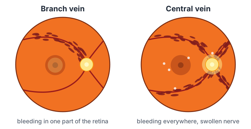
Branch and central
- Branch retinal vein occlusion: a smaller vein is blocked, usually where an artery crosses it and presses it shut. Only part of the retina is affected, and if that part is away from the center, there may be few symptoms.
- Central retinal vein occlusion: the main vein, behind the optic nerve, is blocked. The whole retina is affected, and vision is usually more blurred.
Who is at risk
The same things that harm blood vessels elsewhere in the body:
- High blood pressure, the most important
- Age, usually over 50
- Diabetes, high cholesterol, and smoking
- Glaucoma or high eye pressure, which raise the risk of a central vein blockage in particular
- Less often, conditions that make blood clot more easily, especially in younger people
Because of these links, you will usually be asked to see your primary doctor to check blood pressure, blood sugar and cholesterol. Treating them protects the other eye and the rest of the body.
What it does to vision
- Sudden, painless blurring or a dark area in part of the vision of one eye
- Sometimes it is found on a routine exam with no symptoms
Any sudden change in vision needs a prompt exam; see When to Call the Same Day.
Why vision blurs, and the treatment
Swelling of the macula (macular edema), from fluid leaking out of the congested vessels, is the main reason vision drops. It is treated by a retina specialist:
- Injections into the eye of medicines that stop the leakage. They are the main treatment and usually improve vision. They start about monthly and are spaced out as the swelling settles; many people need them for a year or more.
- Steroid injections or implants, for swelling that does not respond.
- Focal laser, sometimes, for a branch vein blockage.
New, abnormal blood vessels can grow when parts of the retina have lost their blood supply. They can bleed into the gel of the eye or grow over the eye's drain and cause a severe, painful form of glaucoma (neovascular glaucoma). Your doctor watches for them at each visit, most closely in the first months after a central vein blockage. If they appear, they are treated with injections and scatter laser.
What to expect
Many branch vein blockages improve, and most people keep useful vision with treatment. Central vein blockages vary more: some improve well, while others leave lasting blur. The blood clears over months, but the vein itself usually stays partly blocked, so follow-up continues even after vision recovers.
If you have glaucoma
High eye pressure is a risk factor for a central vein blockage, so keeping pressure at target matters for this reason too. After a vein blockage, eye pressure and the drainage angle are checked closely for new vessels. Neovascular glaucoma is treated with injections, laser to the retina, drops, and often a tube shunt or cyclophotocoagulation.
Questions worth asking
- Is it a branch or the central vein, and how much is the macula affected?
- How often will I need injections, and for how long?
- What should I do about my blood pressure and other health checks?
- How will you watch for new blood vessels and glaucoma?