Macular Hole
Published
A macular hole is a small, round break in the very center of the retina, the part used for reading, recognizing faces and seeing detail. It blurs and distorts central vision while side vision stays normal. Most macular holes can be closed with surgery, and the sooner they are repaired, the better the vision tends to be.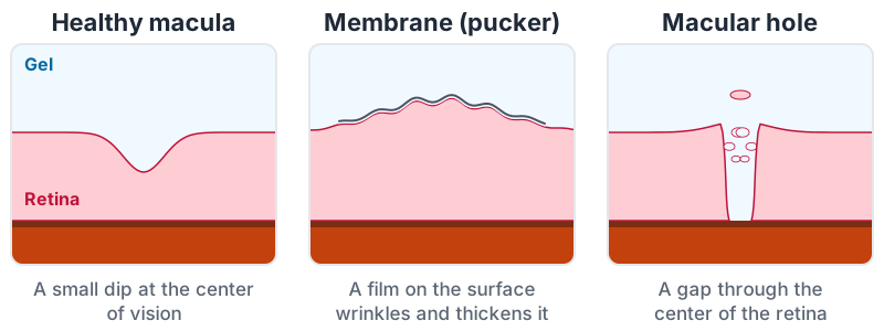
Why it forms
The clear gel that fills the eye, the vitreous, is attached to the retina. With age it shrinks and pulls away (see Flashes and Floaters). If it stays stuck at the center of the macula as it pulls, it can tug the thin center open. This is why macular holes are most common after 60, and more common in women. Less often a hole follows an eye injury, very high nearsightedness, or swelling of the macula.
A macular hole is a different condition from macular degeneration, although both affect central vision.
What it does to vision
- Blurred or distorted central vision in one eye, often first noticed when the other eye is covered
- Straight lines that bend inward toward the center, or a small missing patch in the middle of words or faces
- Side vision is not affected, so it does not cause total blindness
It usually develops over weeks to months. It is painless.
How it is found
A dilated exam shows the hole, and an OCT scan, a quick, painless picture of the layers of the retina, confirms it and measures its size. The scan also shows whether the gel is still pulling on the edges. Because both eyes share the same risk factors, the other eye is checked too; the chance of a hole in the second eye is small but real.
Treatment
Very early holes, where the gel is still pulling but the retina has not fully opened, are sometimes watched, because some release on their own. A full-thickness hole rarely closes without treatment, and the standard treatment is surgery by a retina specialist:
- A vitrectomy removes the gel and its pull on the macula. The surgeon often peels the very thin inner layer of the retina around the hole so its edges can come together.
- The eye is filled with a gas bubble, which presses the edges of the hole together while they heal.
- Head positioning helps the bubble do its job. Many surgeons ask for face-down or a set position for several days; others ask for less. Your surgeon will explain what is needed for your eye.
- Flying and high altitude: while there is gas in the eye there is often a period when you should not fly or travel to high altitude, because the bubble expands as air pressure falls. Tell any doctor who gives you an anesthetic that you have gas in your eye.
Most holes close with one operation. Vision improves gradually over months as the center of the retina reorganizes. Smaller holes, and holes repaired soon after they appear, tend to recover best. Most eyes that still have their natural lens develop a cataract within a year or two of a vitrectomy, and cataract surgery then works well.
Checking at home
Covering one eye at a time and looking at a straight line, or an Amsler grid, is a simple way to notice a change in either eye. A new distortion or blank spot should be reported promptly.
Questions worth asking
- Is the hole full thickness, and how large is it?
- Is surgery recommended now, and what vision might I expect afterward?
- What positioning will I need, and for how long?
- When can I fly, drive and go back to work?
- How likely is my other eye to be affected?