Glaucoma Testing
A glaucoma visit involves more measurements than a routine eye exam, and some of them are repeated at every visit for years. That is not because anything is wrong; it is because glaucoma is diagnosed and followed by looking for change over time. Each test below gives a different view of the same question: is the optic nerve stable?
Eye pressure (tonometry)
The familiar "puff of air" is one way to measure pressure, but in a glaucoma clinic the standard is applanation tonometry: a numbing drop with a yellow dye is placed in the eye and a small blue-lit probe gently touches the cornea. It takes a few seconds and does not hurt. Some clinics also use a handheld device (iCare) that taps the cornea so lightly that no numbing drop is needed.
Pressure varies through the day, often highest in the early morning, so your doctor may want readings at different times of day before changing treatment. Your target pressure is individual, set by how much damage is present and how fast it has progressed.
Corneal thickness (pachymetry)
A quick ultrasound or optical measurement of how thick the cornea is, usually done once. A thick cornea makes pressure readings come out higher than the true pressure and a thin one makes them read lower, so this number helps interpret every pressure reading that follows. A thin cornea is also, on its own, a risk factor for glaucoma.
Looking at the optic nerve
After dilating drops, your doctor examines the optic nerve directly with a lens at the slit lamp. The nerve has a central pale cup and a surrounding pink rim of nerve fibers. In glaucoma the cup enlarges as the rim thins. Your doctor notes the "cup-to-disc ratio," any notching of the rim, and small hemorrhages at the disc edge. Photographs of the nerve may be taken for comparison over the years.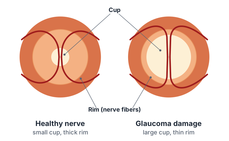
OCT imaging
Optical coherence tomography (OCT) is a painless scan that takes about a minute per eye. You rest your chin on the machine and look at a target while it measures, to within a few thousandths of a millimeter, the thickness of the nerve fiber layer around the optic nerve and the ganglion cell layer in the macula. The report shows your measurements in green (normal), yellow (borderline) or red (thin) against people your age, and, more importantly, tracks your own numbers from visit to visit. OCT often detects thinning before any change shows on the visual field.
Visual field test (perimetry)
This is the test most people remember. You sit at a bowl-shaped machine, cover one eye, stare at a central light, and press a button each time you see a faint flash somewhere in your side vision. It takes about five to seven minutes per eye.
A few things to know:
- It is supposed to be hard. The machine deliberately shows lights at the edge of what you can see. Missing many of them is normal. Keep looking at the center and press when you think you saw something.
- Don't chase the lights. Looking around for the flashes makes the test unreliable. The machine tracks this.
- Results improve with practice. The first field is often worse than later ones simply because the test is unfamiliar. Your doctor may repeat it before relying on it.
- Blink normally and ask for a pause if your eye gets tired; the operator can stop and restart.
The printout maps where sensitivity is reduced. Glaucoma produces characteristic patterns, such as an arc-shaped defect above or below the center or a "nasal step." The software also compares each test to your previous ones and estimates whether the field is stable or getting worse, and how fast.
Gonioscopy
A special mirrored contact lens is placed on the numbed eye at the slit lamp so the doctor can see the drainage angle, which is hidden from direct view. It shows whether the angle is open or narrow, whether it has pigment or other material clogging it, and whether there is scarring or abnormal blood vessels. It is done at the first visit and repeated periodically.
How often tests are repeated
Early on, your doctor may want OCT and visual fields two or three times in the first year to establish a reliable baseline, because detecting slow change requires several good tests. Once stable, many patients have pressure checked every three to six months and imaging and fields once or twice a year. More frequent testing does not mean the disease is worse; it usually means the doctor wants a clearer picture.
Questions worth asking
- What is my target pressure, and what was it today?
- Is my visual field stable compared with last year?
- Is my OCT stable?
- Is there anything in my results that would change treatment?