Epiretinal Membrane (Macular Pucker)
Published
An epiretinal membrane is a thin, see-through film that grows on the surface of the macula, the small central part of the retina used for reading and fine detail. It is also called a macular pucker, because as the film tightens it wrinkles the retina beneath it, like plastic wrap shrinking over a surface. It is common, especially after 50, and many membranes are mild and never need treatment.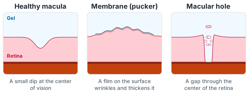
Why it forms
- The gel inside the eye pulling away with age (a posterior vitreous detachment) is the most common setting. Cells left on the surface of the retina grow into a thin sheet.
- After a retinal tear or detachment, or retinal surgery
- After inflammation inside the eye, or blood vessel disease of the retina such as diabetic eye disease or a vein blockage
- Sometimes with no cause found
It is not caused by anything you did, and it is not cancer. It usually affects one eye, though it can occur in both.
What it does to vision
Many membranes cause no symptoms and are found on a routine exam or scan. When they do:
- Distortion: straight lines, such as door frames or lines of text, look wavy or bent
- Blurred central vision, with side vision unaffected
- Objects looking a different size in one eye than the other
- Double vision in one eye, in some people
The changes usually come on slowly and then often stay stable for years. A sudden change in vision is not typical and should be checked promptly.
How it is found
At a dilated exam the membrane can look like a glistening sheen over the macula. An OCT scan, a painless picture of the layers of the retina that takes a few seconds, shows it clearly and measures how much the retina is thickened. Checking each eye on an Amsler grid at home helps you notice whether the distortion is changing.
Treatment
Watching is right for most people: if vision is good and the distortion does not bother you, there is no need to treat. No drops or pills remove a membrane, and glasses cannot correct the distortion.
Surgery is considered when the blur or distortion gets in the way of reading, driving or work. It is done by a retina specialist:
- A vitrectomy removes the gel inside the eye, and the surgeon then peels the membrane off the surface of the retina with fine forceps.
- It is an outpatient operation, usually under a numbing injection with sedation.
- Afterward, vision improves slowly over several months as the retina flattens. Distortion usually lessens a great deal, though it may not disappear completely. The longer and worse the distortion before surgery, the less complete the recovery tends to be.
- Most eyes that still have their natural lens develop a cataract within a year or two of a vitrectomy, and cataract surgery then works well.
- A membrane rarely grows back.
If you have glaucoma
A membrane can make the OCT scans and visual fields used to follow glaucoma harder to read, so your eye doctors may compare tests with this in mind. Eye pressure is checked closely after any retinal surgery.
Questions worth asking
- How much of my blurred vision is from the membrane, and how much from anything else, such as a cataract?
- Is it changing on my scans?
- At what point would surgery be worth it for me?
- What would recovery look like for my eye?