Diabetic Eye Disease
Diabetes can damage the small blood vessels of the retina. This is diabetic retinopathy, and it is a leading cause of vision loss in working-age adults. It causes no symptoms in its early stages, when treatment works best, so regular dilated eye exams are the way it is caught. Most serious vision loss from diabetes can be prevented.
Press Play to see the stages.
The animation needs JavaScript. The pictures and the explanation below cover the same thing.
How diabetes affects the retina
High blood sugar, over years, weakens the walls of the retina's small blood vessels.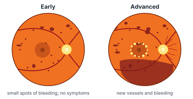
- Non-proliferative retinopathy. The vessels bulge, leak and close off. The doctor sees dots of bleeding and yellow deposits. Vision is usually normal.
- Diabetic macular edema. Leaking near the macula makes it swell, which blurs central vision. This can happen at any stage and is the most common way diabetes affects sight.
- Proliferative retinopathy. The starved retina grows new blood vessels. They are fragile and can bleed into the gel that fills the eye, or scar and pull the retina off. This stage threatens severe vision loss.
Diabetes also brings cataract on earlier, makes glaucoma more likely, and can blur vision for days or weeks when blood sugar swings, by changing the focus of the lens.
Symptoms
Often none, even with significant disease. When there are symptoms:
- Blurred or fluctuating vision
- New floaters, a shower of spots, or a red or dark haze, from bleeding
- Dark or empty areas in vision
A sudden shower of floaters or loss of vision needs a same-day call.
How often to be examined
- Type 2 diabetes: at diagnosis, then every year.
- Type 1 diabetes: starting within five years of diagnosis, then every year.
- Pregnancy with diabetes: early in the pregnancy, and more often during it.
- More often once retinopathy is present, as your doctor advises.
The exam uses drops to dilate the pupils, and often a photograph or a scan of the macula (OCT). A photo screening without dilation is a reasonable check when the eyes have been normal, but it does not replace a full exam once changes are found.
What you can do
Good control slows every stage of this, and it is never too late to benefit.
- Blood sugar. Each improvement in the A1c lowers the risk.
- Blood pressure and cholesterol, which matter nearly as much.
- Do not smoke.
- Keep your eye appointments, even when you see well.
If your blood sugar is brought down very quickly after a long period of poor control, retinopathy can worsen for a time, so your eye doctor should know about big changes in treatment.
Treatment
- Injections into the eye of medicines that stop leakage and make new vessels shrink. They are the main treatment for macular edema and are also used for proliferative disease. They are repeated, often monthly at first.
- Laser. Scatter laser (PRP) to the outer retina makes new vessels regress and has protected vision for decades. Focal laser treats some leaking spots near the macula.
- Steroid injections or implants for swelling that does not respond.
- Surgery (vitrectomy) to clear blood that does not go away, or to release scar tissue pulling on the retina.
These are done by a retina specialist. Your eye doctor will refer you when retinopathy reaches the stage where treatment helps.
Questions worth asking
- Do I have any retinopathy, and what stage?
- Is my macula swollen?
- How soon should I be seen again?
- What A1c and blood pressure should I aim for to protect my eyes?